Head and neck cancers: operating through the mouth without facial incisions

Bathsheba Huruy

Publié il y a 0 jours

25.09.2026

Partager
At Lausanne University Hospital (CHUV), the new single-port Da Vinci robotic system allows surgeons to operate on certain areas of the throat directly through the mouth. In Vivo followed one such procedure.

Reaching a tumour deep in the throat – in the oropharynx, around the tonsils and the base of the tongue – can require major surgery, sometimes even involving cutting through the jaw to access the area being operated on. With the single-port Da Vinci robotic system, some of these procedures can now be performed directly through the mouth. At CHUV, where the system has been in use since 2026, two surgeons work in tandem: Professor Christian Simon, head of the Department of ENT and Head and Neck Surgery, controls the robot’s instruments from a console in the operating theatre, while Dr Karma Lambercy, a senior physician in the same department, assists at the patient’s side.

The main advantage of the technique in head and neck surgery lies in this access through the mouth. Known as a “transoral” approach, it helps limit the effects of surgery: no scarring on the face or neck, shorter hospital stays, and better preservation of functions such as swallowing and speech. The technique is not appropriate for every tumour, however. It is intended for relatively small lesions that can be reached through the mouth. For many patients, the alternative is not conventional surgery but radiotherapy, with or without chemotherapy. According to Professor Christian Simon, 20–30% of oncological surgery in the specialty could benefit from robotic assistance. Last July, In Vivo followed one such operation step by step, as surgeons removed a suspicious mass from a patient’s throat.
1/ Gaining access to the throat

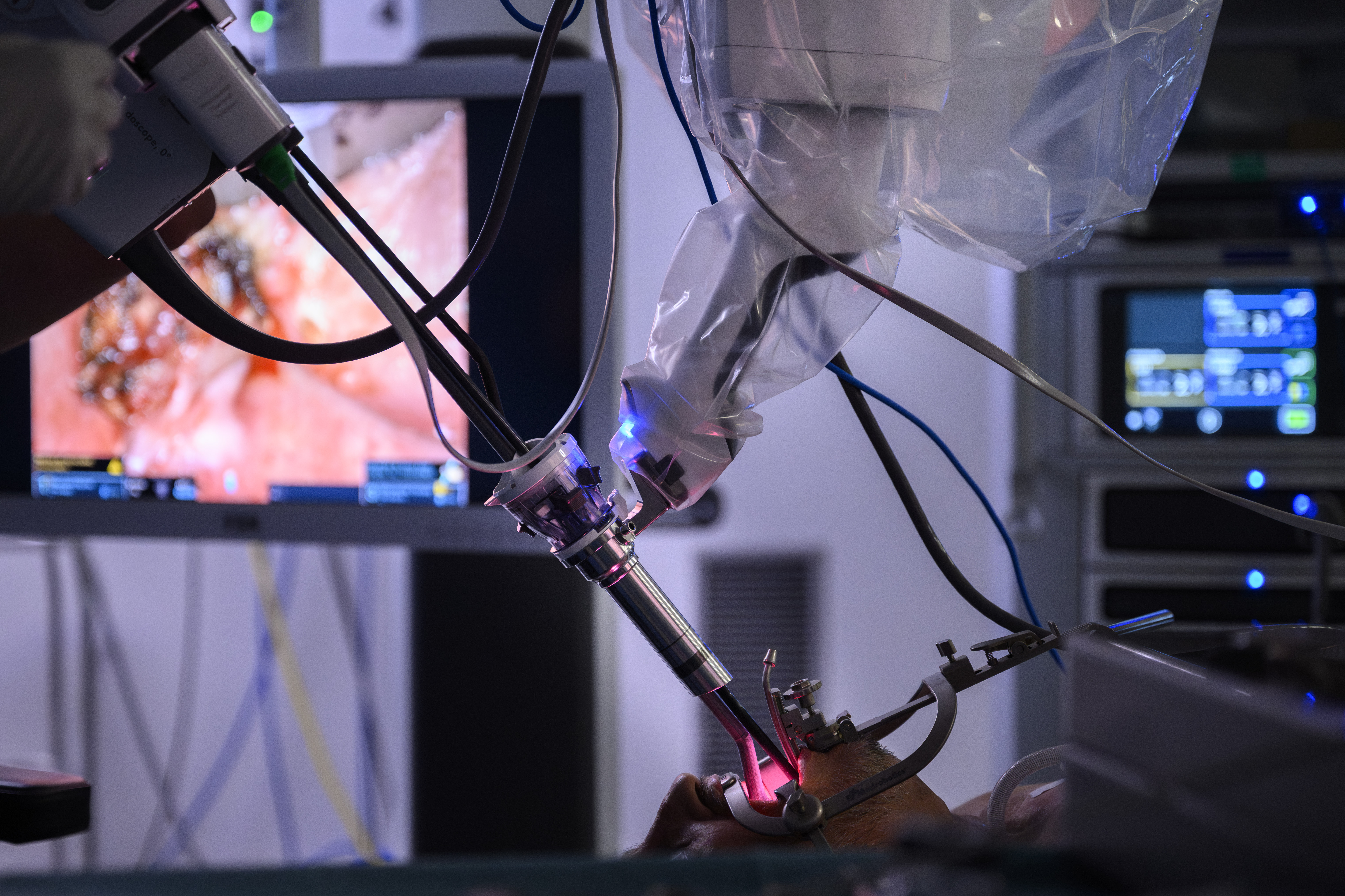
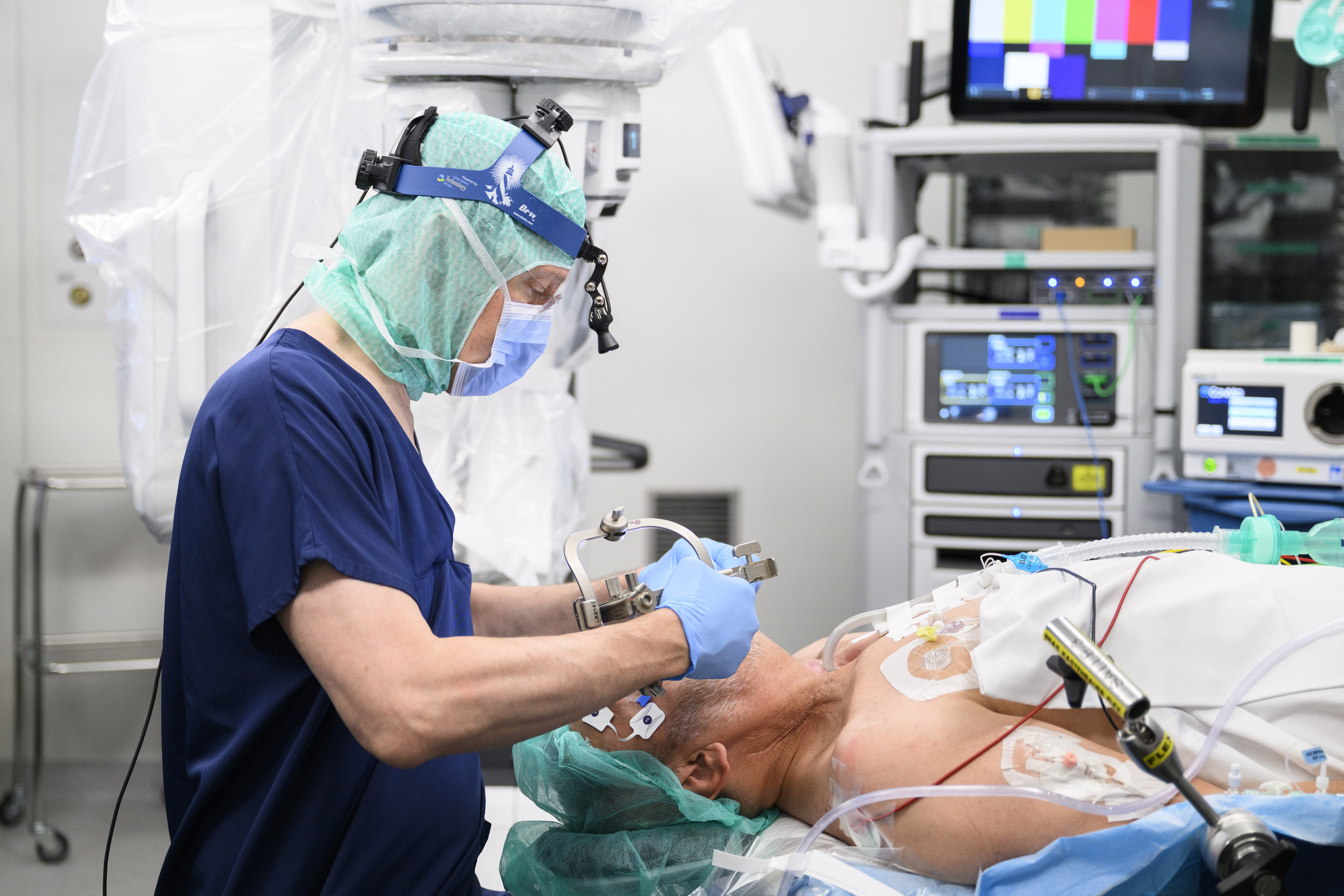
The patient is placed under anaesthesia and positioned on his back, with his head slightly tilted backwards to improve access through the mouth. A retractor holds the mouth open and exposes the part of the throat the surgeons need to reach.

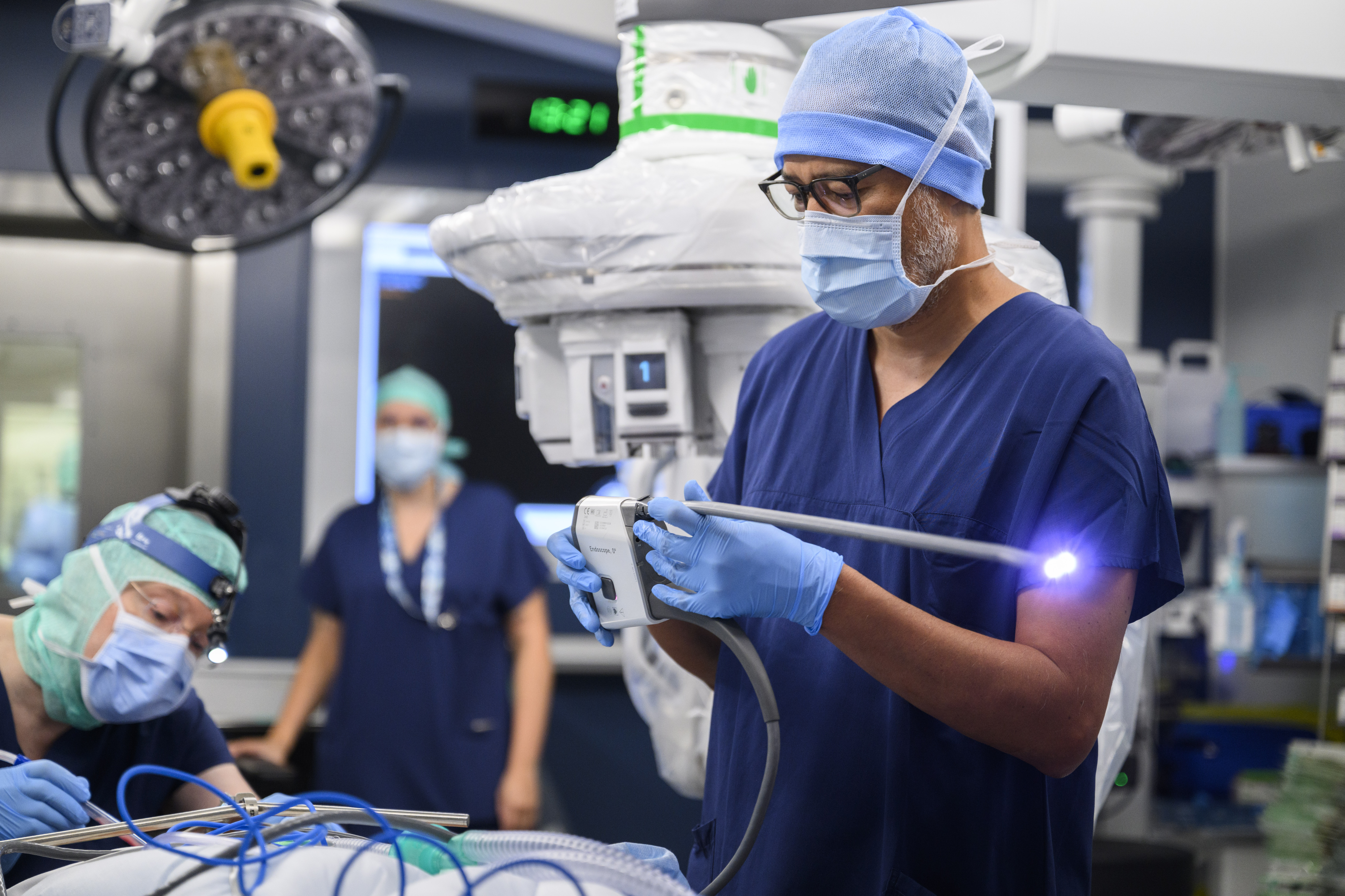
2/ Positioning the camera

Karma Lambercy attaches the camera to the robot’s arm. Its flexible design allows it to be repositioned during the procedure, giving different views of the surgical site.

3/ Bringing the robot into position

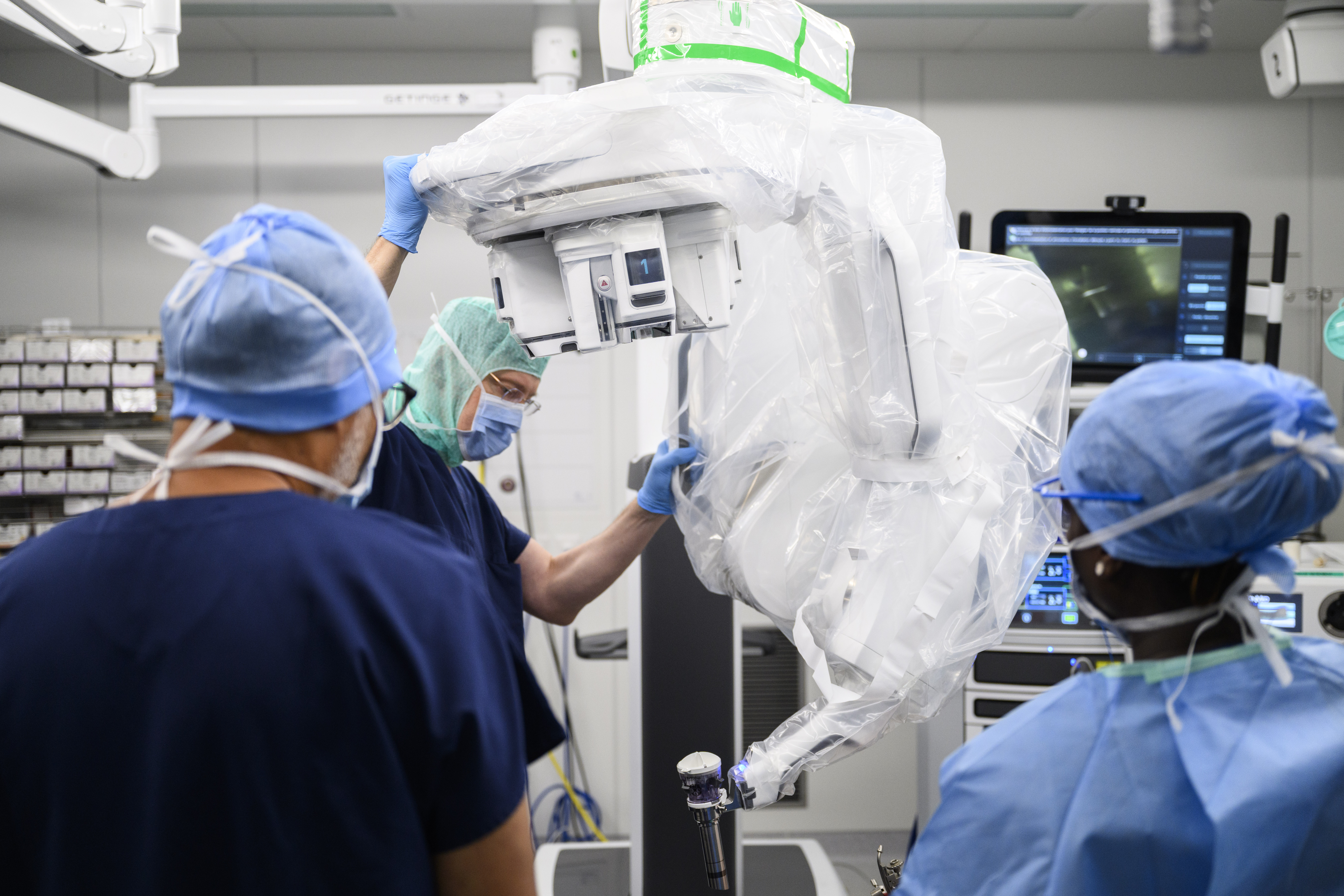
Once the patient is prepared and the camera is in place, the “docking” stage begins. This involves bringing the robot up to the operating table and positioning its arm above the patient’s mouth.

4/ Accessing the throat

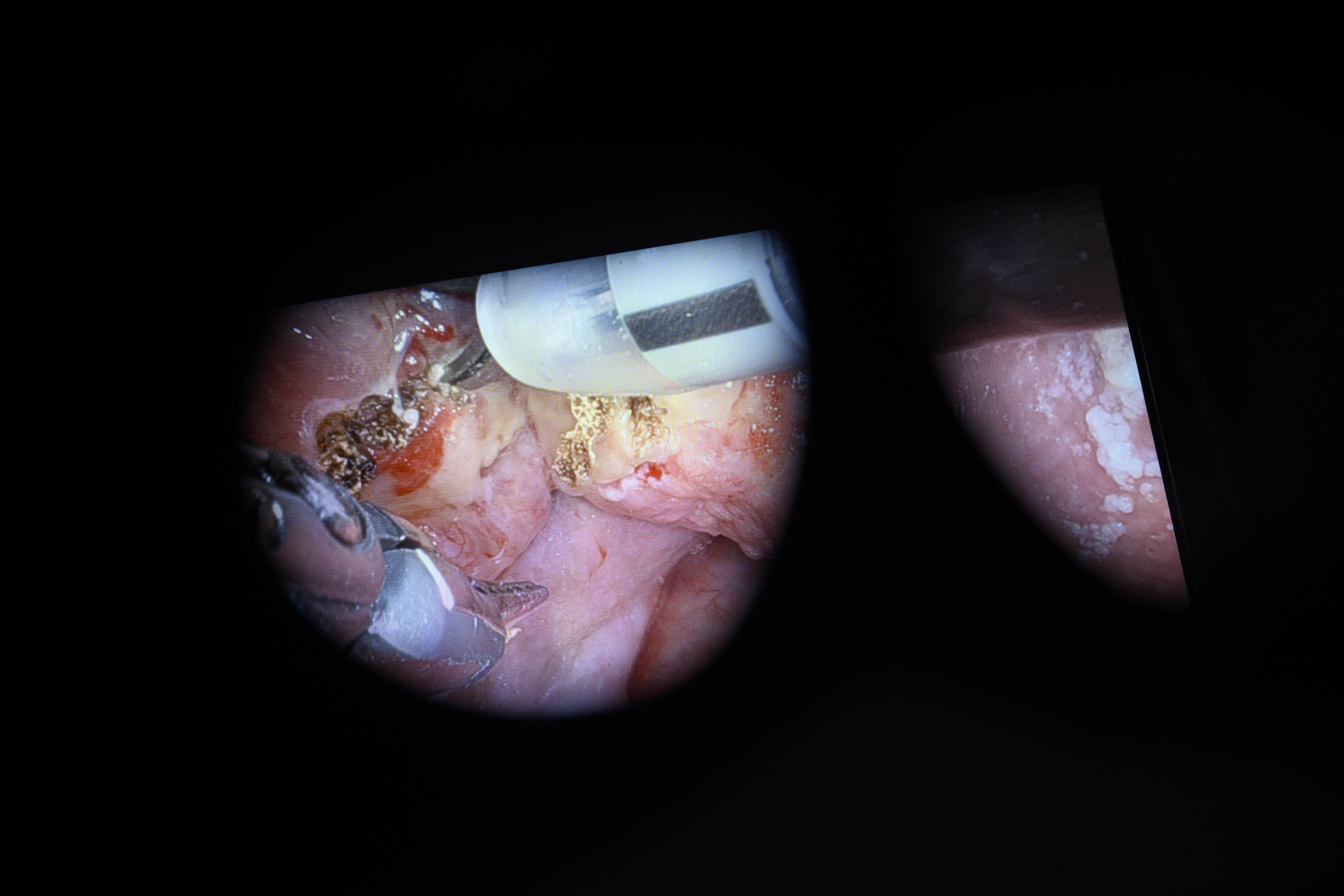
The camera, scissors and Maryland forceps are inserted through the patient’s mouth and advanced to the surgical site. This approach makes it possible to reach the throat without opening the neck or cutting through the jaw.

5/ Operating from a console

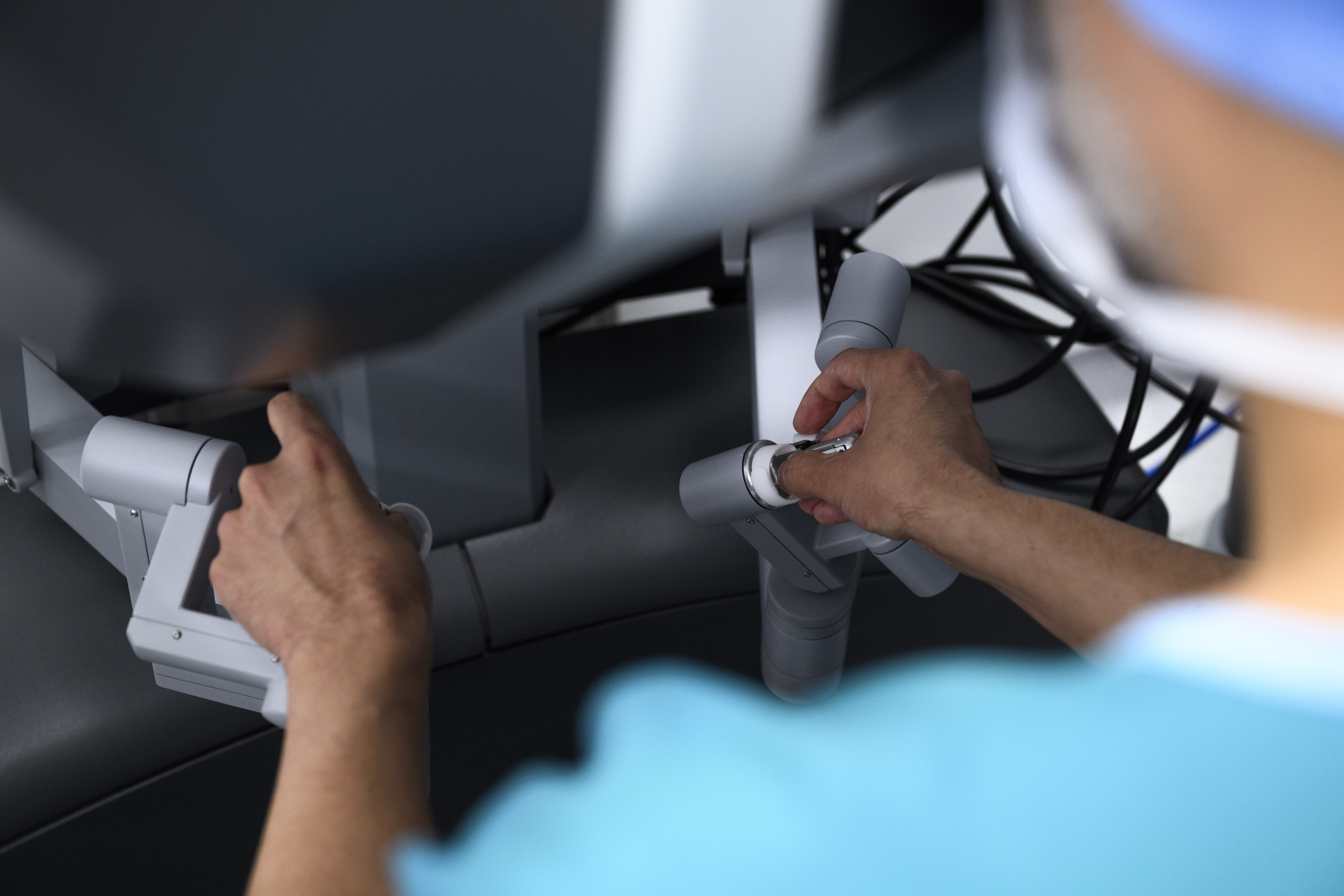
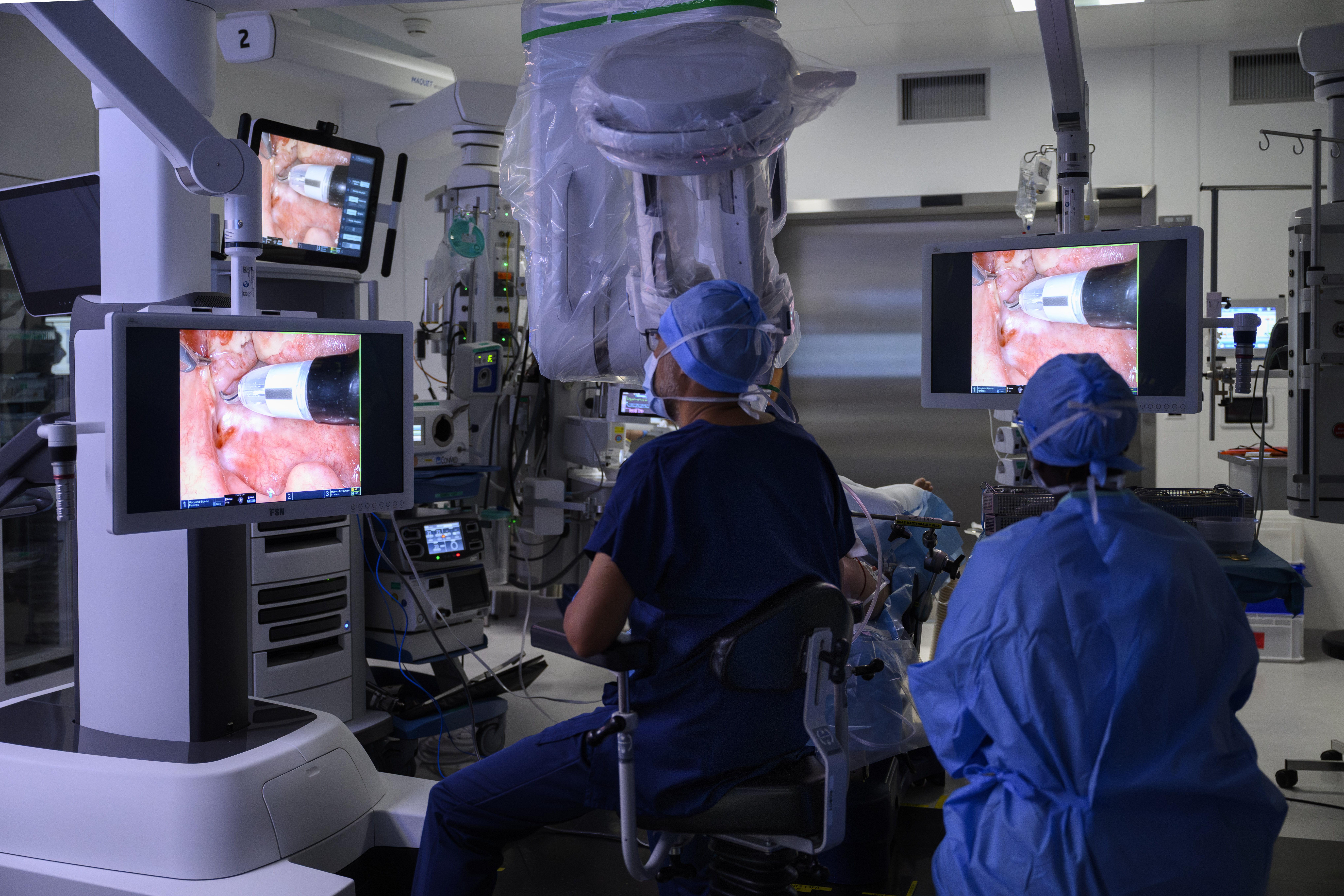
A few metres from the patient, Christian Simon operates from a console displaying highly magnified 3D images of the surgical site. Using hand controls and foot pedals, he directs the movements of the camera, scissors and Maryland forceps inserted into the throat.

6/ Working in tandem

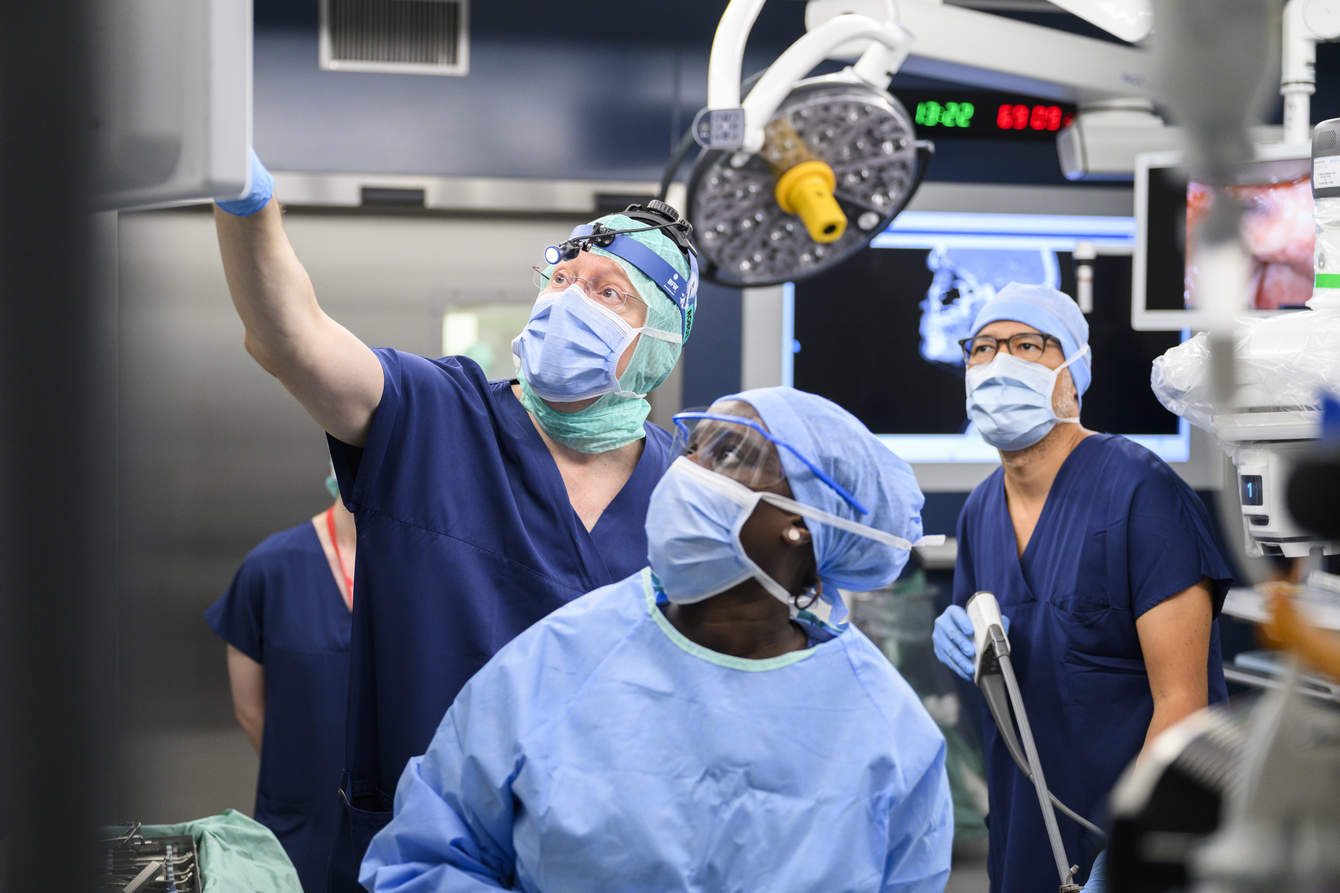
The procedure involves two surgeons with complementary roles. While Christian Simon controls the robot from the console, Karma Lambercy works beside the patient’s head. Guided by the camera images, he suctions blood and secretions, applies clips to seal blood vessels when necessary, and repositions the tongue to improve access to the surgical site.

7/ Cutting and controlling bleeding

The instruments inserted into the throat include scissors and Maryland forceps. An electric current passes through the scissors, allowing them to cut tissue while coagulating small blood vessels and thereby limiting bleeding as the mass is removed.

8/ Analysing the tissue

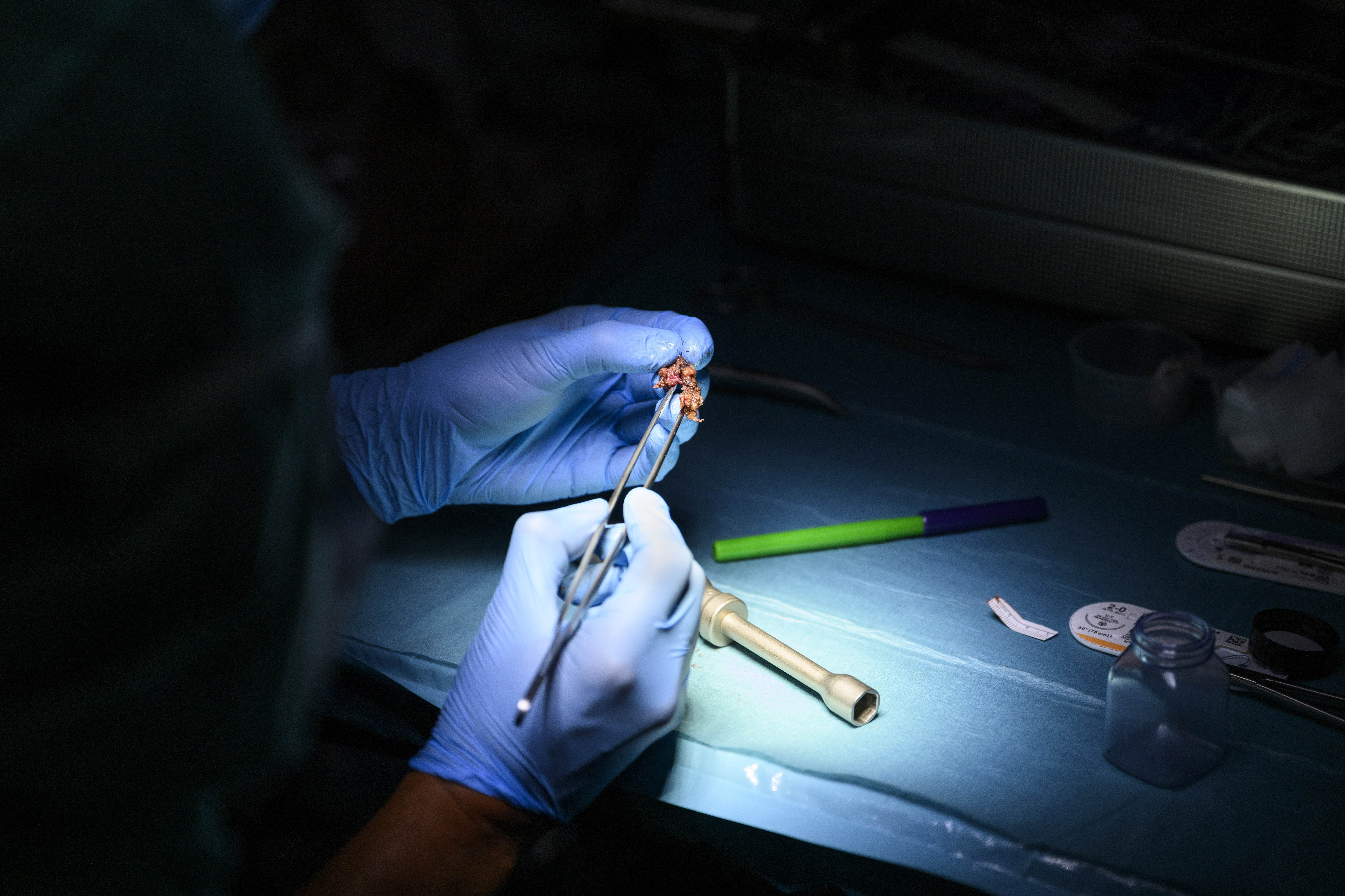
The tissue removed during surgery is sent to the pathology team. An initial examination carried out during the procedure helps guide the surgery in real time. The final diagnosis, however, is based on a full analysis of the samples, with results available only a few days later.