Beware of fake scientific images
Publié il y a 10 mois
24.03.2025
Partager

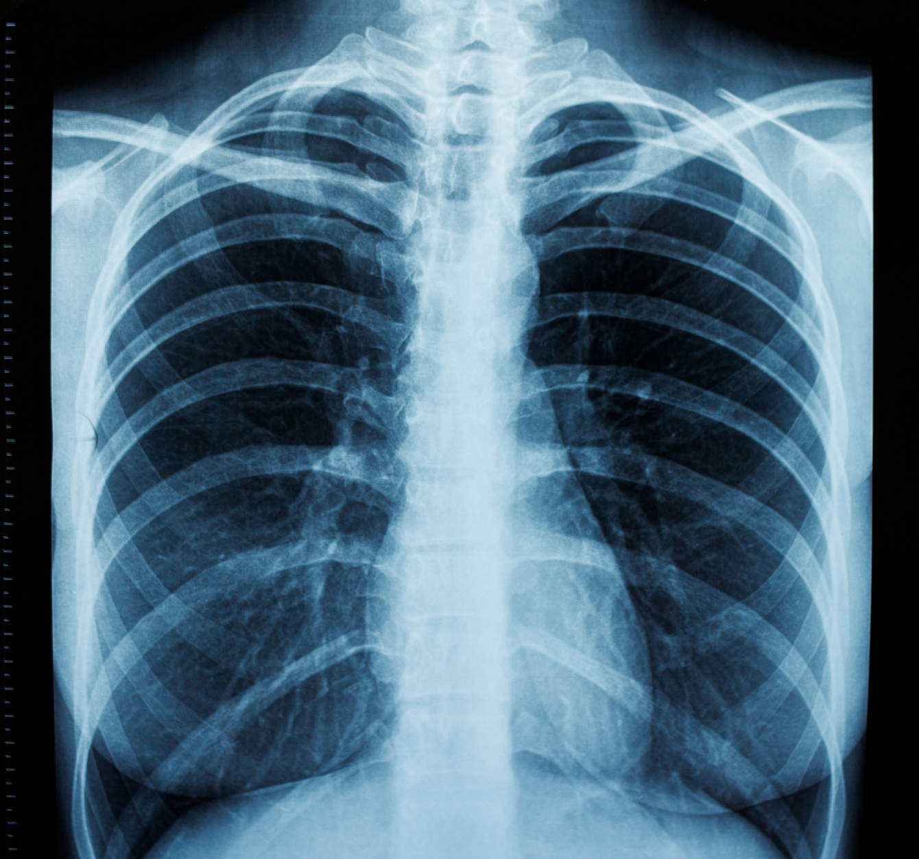
With the rise of artificial intelligence (AI), academic plagiarism has become even more problematic. A study published in 2022 in the European Journal of Radiology indicated that among the 219 authors who recently published in the top twelve radiology journals, 27.4% had either witnessed or suspected fraud by their colleagues.
The use of software to detect plagiarism in scientific texts is now widespread. On the other hand, programs dedicated to images are not as developed or used. With the deployment of AI, falsification and plagiarism of scientific images are becoming a major challenge. This is what radiologists Thomas Saliba and David Rotzinger of the CHUV’s Department of Radiodiagnosis and Interventional Radiology argue in an article published earlier this year in the journal European Radiology. “The majority of people think that the scientific community is rigorous and honest, and even if that were not the case, it would be difficult to falsify images conclusively, says Saliba. For some years now, there has been no need for special technical knowledge to falsify images convincingly. Sometimes they become almost impossible to detect.”
Radiology at risk
In 2023 alone, more than 10,000 articles were removed for plagiarism and fraud, reports the journal Nature. “A tendency underestimated by the scientific community”, according to Thomas Saliba, who also explains it by the immense pressure on researchers to publish as much as possible. Radiology relies heavily on digital images, so it’s a higher risk speciality, says the specialist.
“Evidence validating a new technology or technique can be potentially falsified, which can be very dangerous,” warns David Rotzinger, who is also a teacher at UNIL. For example, doctors at the University of Oxford claimed to have developed a technique to differentiate lesions without using Gadolinium, a rare metal used as a contrast agent in MRI. But, it turned out that the images and therefore the results were false. “If the fraud had not been detected, patients could have received a false diagnosis and therefore potentially inappropriate treatment.”
Radiology is not the only field that is affected. Images are used in all medical specialities, says Rotzinger. “Often, we compare what we see in our clinical examinations with examples in the literature. If we can no longer trust what is in books and papers, our work becomes much more difficult.”
Thomas Saliba and David Rotzinger believe that image manipulation must be detected in the same way as textual plagiarism. Some software companies, such as Proofig AI or Imagetwin.ai, already offer this type of service. However, both researchers consider that AI is comparatively neglected as a tool for scanning and detecting falsified images submitted to journals.
In their work as reviewers, the duo even found images taken from other sources that the authors of the submitted articles tried to pass off as their own after changing their orientation. In such cases, a simple reverse image search on Google using the submitted article’s image can show exactly from which publication it was taken, says Saliba. “This method - extremely simple but effective - would be a good basis for an algorithm to detect and prevent all forms of plagiarism.”
To go further
Thomas Saliba and David Rotzinger's article “Figure plagiarism and manipulation, an under-recognised problem in academia” was published in the journal European Radiology in February.
Link to article:
https://link.springer.com/article/10.1007/s00330-025-11426-2


![[Translate to Anglais:] IMAGE: CHUV](https://www.invivomagazine.ch/fileadmin/_processed_/3/f/csm_ROTZINGER_David_20210202_017_0bf35cb784.jpg)

![[Translate to Anglais:] CRÉDIT: Gilles Weber](https://www.invivomagazine.ch/fileadmin/_processed_/1/a/csm_54621_25_DAL_Robot_MAKO_3930_af7417e685.jpg)